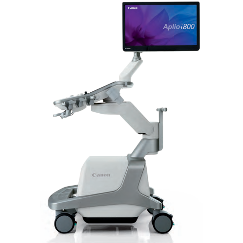
The Canon Aplio i800 is a high-end ultrasound system designed to deliver exceptional clinical accuracy and optimal productivity. With its ultra-wideband transducers, imaging technologies such as SMI and iDMS, and intuitive interface, it provides clear and detailed visualisation of anatomical structures, even in the most complex patients. Lightweight, easy to handle and equipped with intelligent automation tools, the Aplio i800 adapts to all clinical environments, from general diagnostics to specialised examinations, while ensuring consistent image quality and smooth navigation.
Aplio i800 Ultrasound Scanner - Fair Condition 2018


Offer Details
Accessories :
Price without probes, contact our team for probe options (for additional price)
Description
Aplio i800: Precision imaging for reliable diagnostics
The Canon Aplio i800 is designed to meet the highest standards in medical diagnostics. Its iBeam technology, combined with increased processing power, delivers exceptional image clarity and improved penetration, even in patients with high body mass index. With iDMS (Dynamic Micro-Slice) technology, slice thickness is electronically refined to reveal more detail at all depths. Ultra-wideband transducers cover an extended frequency range, equivalent to two conventional probes, ensuring superior sensitivity and resolution in both near and far fields.
Cutting-edge technologies for optimal visualisation
The Aplio i800 incorporates advanced imaging technologies such as ApliPure+ and Precision+, which reduce speckle noise, improve contrast and enhance the definition of anatomical contours. Adaptive slice thickness control simultaneously optimises resolution and sensitivity in each imaging mode. SMI (Superb Micro-vascular Imaging) technology revolutionises Doppler imaging by visualising low-velocity microvascular flow that was previously invisible. It enables accurate assessment of organ and lesion microvascularisation with unparalleled detail.
The system also supports CEUS (Contrast-Enhanced Ultrasound) imaging and shear wave elastography, with propagation mapping and dispersion measurement, for quantitative assessment of tissue stiffness and viscosity. ATI (Attenuation Imaging) allows the attenuation coefficient in the liver to be measured, excluding structures such as vessels and calcifications for robust results.
Ergonomics, connectivity and intelligent automation
The Aplio i800 has been designed with user comfort in mind: lighter and more compact than its predecessors, it features a screen that can be adjusted over 36 cm, an articulated arm and a sliding side panel. Its touchscreen interface with three interactive zones makes navigation and access to functions easy. The system can be controlled remotely via a wireless tablet, ideal for mobile or sterile environments.
Automation tools such as Real-time Quick Scan and integrated multi-parameter measurements speed up examinations while ensuring consistent quality. The Smart Fusion feature allows ultrasound images to be merged in real time with previously acquired data (CT, PET-CT, MRI), facilitating lesion identification and navigation in complex anatomies. Finally, improved visualisation of biopsy needles thanks to BEAM technology enhances the safety and accuracy of interventional procedures.
Features
- High-resolution imaging with noise reduction
- Visualisation of low-velocity microvascular flow (SMI)
- Adaptive slice thickness control
- Tissue harmonic imaging
- Quantitative measurement of tissue elasticity and viscosity
- Multiparametric liver assessment
- Intuitive navigation with contextual interface
- Automation of settings and measurements
- Compatibility with multi-angle needle guides
- Smart Navigation function for guided biopsies
Technical Details
- Ultra-wideband transducers up to 33 MHz
- High-processing power iBeam technology
- iDMS technology for electronic slice thickness refinement
- Doppler modes: colour, power, SMI
- CEUS and ATI imaging
- Shear wave elastography with propagation mapping
- Quad mode: simultaneous display of 4 modes
- 36 cm adjustable screen, articulated arm
- Touchscreen interface with 3 interactive zones
- Smart Fusion function (fusion with CT, MRI, PET-CT)
- Needle visualisation with BEAM technology
- Integrated raw data for lossless post-processing
Compatible Accessories
- Specialised transducers:
- Thin convex for intercostal access
- High-frequency linear (up to 33 MHz)
- Volume matrix for 3D imaging
- Hockey stick for small structures
- Needle guides:
- Multi-angle or free-angle
- Support mounting or direct mounting
- Wireless tablet for remote control
- Biopsy attachment with selectable puncture angle
- Secondary console for dual operator