• Medical Diagnostic Equipment
  • Surgical and Therapeutic Equipment
  • Medical Furniture
  • Laboratory Equipment
  • Cleaning and Sterilization

Confocal Retinal Angiography Device HRA 2 - Good Condition


The HRA 2 is a confocal scanning laser ophthalmic diagnostic system designed for fluorescein and indocyanine green angiography. Providing high-precision digital retinal imaging, it enables the acquisition of single-layer, time-sequence, or three-dimensional images. Thanks to its multiple laser wavelengths and its ability to perform combined imaging modalities, the HRA 2 stands out as an essential tool for the screening and monitoring of retinal pathologies.

Offer Details

1 Introduction 1.1 Intended use The Heidelberg Retina Angiograph 2 is a diagnostic device for the human eye. Intended use is for the examination of the retina by laser scanning either with or without contrast agent, resp. fluorescence agent). The purpose of the angiography testing is to assist in the diagno- sis of eye diseases. The diagnostic result shall provide help to the physician for adequate therapy. 1.2 The Heidelberg Retina Angiograph 2 The Heidelberg Retina Angiograph 2 is a confocal laser scanning system for digital fluorescein and indocyanine green angiography. Fluorescein and indocyanine green angiography can be per- formed either separately or simultaneously. For acquisition of the digital confocal images, a laser beam with a wavelength suitable to excite the fluorescent dye is focused on the retina. The laser beam is deflected periodically by means of oscillating mirrors to sequentially scan a two-dimensional sec- tion of the retina. The intensity of the emitted fluo- rescent light at each point is measured with a light sensitive detector. In a confocal optical system, light emitted outside of the adjusted focal plane is sup- pressed. This suppression increases with the distance from the focal plane, and results in a high contrast image. Furthermore (especially during indocyanine green angiography), the confocal optical system al- lows the user to acquire three-dimensional images layer by layer, which is comparable to laser scanning tomography. The laser sources contained in the Heidelberg Retina Angiograph 2 emit laser light of three different wave- lengths: for fluorescein angiography, the blue line of a solid state laser (488 nm) is used to excite the fluo- rescein. A barrier filter at 500 nm edge wavelength separates excitation and fluorescent light. The same wavelength (but without barrier filter) is used to create red free reflectance images. For indocya- nine green angiography, a diode laser at 795 nm wavelength is used to excite the dye, while a bar- rier filter at 810 nm edge wavelength separates excitation and fluorescent light. A second diode laser at 830 nm wavelength serves to produce infrared reflectance images. Several image acquisition modes are provided, i.e. pure fluorescein angiography, pure indocya- nine green angiography, and simultaneous fluorescein / indocyanine green angiography. In the simultaneous mode, each image line is scanned twice. During the first scan, fluorescein is excited and its fluorescence is measured; during the subsequent second scan, indocyanine green is excited and its fluorescence is measured. In every acquisition mode, individual images, time resolved im- age sequences, or a focal series of images (layered, three-dimensional images) can be acquired. The images are immediately digitized with a resolution between 384 x 384 and 1536 x 1536 pix- els. The digitized images are stored in the computer's main memory and then saved on the com- puter's hard disk. The size of the field of view can be set to 15 x 15 degrees, 20 x 20 degrees, or 30 x 30 degree

Description

Confocal Scanning Retinal Imaging: The Excellence of Heidelberg’s HRA 2

The HRA 2 by Heidelberg is an ophthalmic diagnostic system based on confocal scanning laser technology. It captures high-contrast retinal images with or without contrast agents, facilitating the detection of both vascular and structural abnormalities. Using a two-dimensional laser scanning mechanism that focuses light on specific retinal areas, it generates remarkably contrasted images. The confocal rejection of out-of-focus light ensures clear imaging by eliminating stray signals, delivering optimal image quality even in deeper layers—particularly valuable in indocyanine green angiography.

The device features three distinct laser sources: a 488 nm semiconductor laser for fluorescein angiography, a 795 nm diode laser for indocyanine green, and an 830 nm diode laser for infrared reflectance imaging. These wavelengths allow for precise mapping of retinal vascularization. In simultaneous imaging mode, the HRA 2 scans each image line twice, capturing both dye signals in parallel. This dual capture capability enhances clinical relevance while streamlining examination protocols.

Images are acquired in high resolution up to 1536 x 1536 pixels, and are immediately digitized and stored on the control computer. The system offers adjustable fields of view—15°, 20°, and 30°—with the ability to create panoramic composites up to 120°. Deep layer explorations, similar to tomography, are also possible through sequential layered image capture.

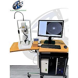
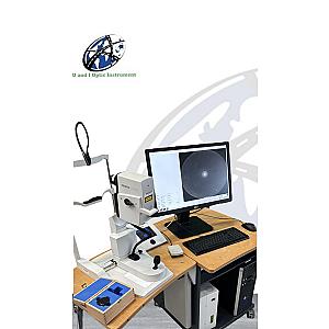
The system includes a laser scanning camera, a touchscreen control panel, a laser module power supply, a computer with dedicated software, and an isolation transformer compliant with IEC 60601-1 standards. Operation is guided via an intuitive interface using a touchscreen, keyboard, or footswitch. Sensor sensitivity, focus, and scanning parameters are finely adjustable to ensure image acquisition tailored to each clinical scenario.

The HRA 2 combines technological precision, ergonomic design, and flexible operation, supporting eye care professionals in their most complex diagnostic challenges.

Features

  • Confocal scanning system for digital angiography
  • Single-layer, time-lapse, or 3D image acquisition
  • Separate or simultaneous modes for fluorescein and indocyanine green angiography
  • Precise filtering of out-of-focus light signals
  • Real-time adjustable sensor sensitivity
  • Ergonomic interfaces: touchscreen, footswitch, keyboard
  • Compatible with Heidelberg Eye Explorer software for analysis and archiving

Technical Details

  • Field of view: 15°, 20°, 30° (composite up to 120°)
  • Focusing range: –12 to +12 diopters, in 0.25 D increments
  • Available resolutions: High resolution: up to 1536 x 1536 pixels
  • Maximum frame rate: High resolution: up to 9 Hz; Fast mode: up to 16 Hz
  • Z-scan depth: up to 8 mm
  • Digital resolution: 5 µm/pixel (HR), 10 µm/pixel (HS)
  • Laser sources: 488 nm (FA), 795 nm (ICGA), 830 nm (IR)
  • Power consumption: 70 W
  • Input voltage: 100–240 V / 50–60 Hz

Compatible Accessories

  • Fluorescein sodium and indocyanine green dyes
  • Corrective lenses for astigmatic patients (>2D)
  • Additional patient headrests and fixation supports
  • External storage devices for archiving (FireWire hard drive)
  • Lens cleaning supplies (acetone and lint-free cotton)
  • Heidelberg software updates and optional add-on modules

Discover more in this category

Logistics

Remma and its partners provide you with serviced and configured equipment tailored to your needs. Receive your product at your facility with standard or express delivery, and an installation option, all through a single point of contact.

Warranty

Our 12/24-month warranty ensures the safety and reliability of your medical equipment throughout its lifespan.

Flexible Financing

Settle your medical equipment in up to 36 installments with our financing service or pay after delivery with Remma Facility.