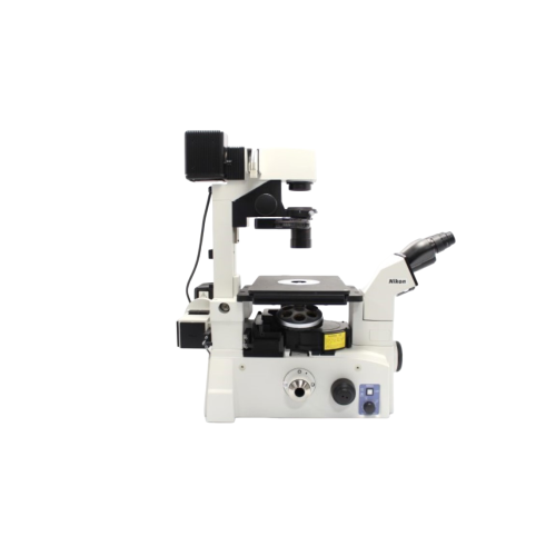
Eclipse TE2000-U: An inverted microscope designed for cutting-edge research
The Nikon Eclipse TE2000-U is an inverted research microscope specially designed to meet the needs of academic and pharmaceutical laboratories working in the fields of cell biology, molecular biology and fluorescence microscopy. Its modular architecture makes it a versatile platform, capable of adapting to technological developments and the specific needs of each user. Unlike traditional upright microscopes, the inverted model allows for optimal observation of living samples in culture dishes or flasks, making it an indispensable tool for real-time imaging.
Optical performance and optimised light transmission
One of the major advantages of the Eclipse TE2000-U is its state-of-the-art optical system, based on Nikon CFI60 technology. This design offers increased working distance and high numerical aperture, enabling sharp, high-contrast images to be captured even in low-light conditions. The internal light path has been optimised to reduce transmission losses and improve fluorescence quality. This provides researchers with accurate and reproducible imaging, which is essential for applications in cytometry, cell tracking and structural biology.
In addition, the mechanical stability of the stand and the precision of the controls ensure reliable focusing and ease of use during prolonged experiments. The optical components are designed to minimise chromatic aberration and maximise colour reproduction, an essential asset for multi-parameter observations.
A scalable platform for laboratory applications
The Nikon Eclipse TE2000-U has been designed as a scalable platform capable of accommodating various add-on modules. It can be equipped with multi-channel fluorescence devices, specialised illuminators and high-resolution digital imaging systems. Its interface is compatible with many Nikon and third-party accessories, allowing complete customisation according to experimental needs.
This flexibility makes it the tool of choice for applications such as confocal imaging, multiphoton microscopy and cellular photomanipulation. Thanks to its ergonomic and robust design, it supports complex configurations while remaining extremely easy to use. Researchers can therefore implement sophisticated protocols without compromising imaging quality.