• Medical Diagnostic Equipment
  • Surgical and Therapeutic Equipment
  • Medical Furniture
  • Laboratory Equipment
  • Cleaning and Sterilization
  • Spare Parts

EpiCam V Portable Veterinary Retinal Camera - Fair Condition

The EpiCam V from epipole is a portable retinal camera designed for veterinary practice, allowing digital colour images of the fundus to be captured through the pupil. With a resolution of up to 10 μm and real-time video capability at 14 frames per second, it facilitates the early detection of retinal micro-abnormalities. Compact and lightweight, it connects directly to a PC, laptop or tablet via USB, with no internal battery to recharge. It also offers the possibility of capturing images of the anterior segment, while ensuring optimal comfort for the animal thanks to safe, continuous lighting without flash.


Offer Details

Options :

EPIPOLE Limited EpiCam V Veterinary Technology EpiCam Vet Camera fundus camera

The Offer Details are provided by the seller and automatically translated.

Description

EpiCam V: High-Precision Retinal Imaging for Veterinary Practice

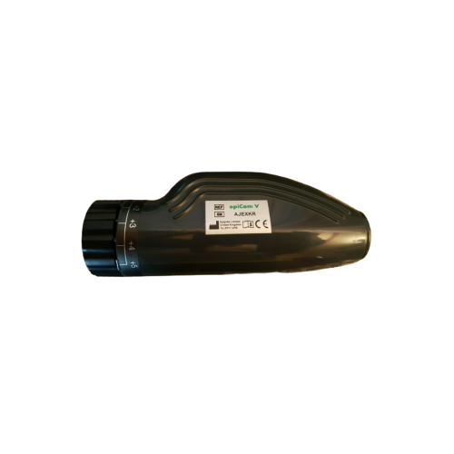
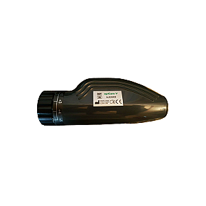
The EpiCam V from epipole is a portable camera specially designed for imaging the fundus of the eye in animals. Used in veterinary ophthalmology, it captures digital colour images of the retina through the pupil without touching the eye, ensuring optimal comfort for the animal. With a resolution of approximately 10 µm, it can distinguish very fine details, such as microaneurysms, facilitating the early detection of eye abnormalities. The device can produce a real-time video stream at 14 frames per second, with the option to freeze and record the image at any time. The wide field of view and the ability to rotate or tilt the device allow for quick exploration of areas of interest in the retina.

Versatile Use and Optimised Ergonomics

Designed for simple and efficient use, the EpiCam V connects directly to a laptop, tablet or PC via USB 3.0 for full resolution of 2592 × 1944 pixels, or via USB 2.0 at 1296 × 972 pixels. Its focus correction range of more than ± 15 diopters and working distance of 13 mm ensure optimal sharpness for different types of eyes, including pupils with a diameter of 4 mm or more. In addition to retinal imaging, the device can be used to capture images of the anterior segment, such as the cornea and iris, by adjusting the distance and lighting. The software allows you to create and manage patient files, capture and review images and videos, adjust settings (brightness, contrast, gamma) and export files in PNG, TIFF or JPEG formats. Integrated telemetry sensors record the position of the device, improving accuracy during the examination.

Safety, Comfort and Durability

The EpiCam V uses low-power continuous LED lighting, classified as Group 1 according to ISO 15004, ensuring safe use for animals' eyes. It is recommended to limit the examination to less than two minutes per eye to reduce any temporary visual after-effects. Compact (64 × 44 × 153 mm) and lightweight (185 g, excluding cable), it is easy to handle and adapts to a variety of clinical environments, including dimly lit rooms to facilitate pupil dilation. Its robust design and durable housing ensure a long service life. Maintenance is simple: the lens can be cleaned with a blower or microfibre cloth, avoiding any products that could damage the optical surface. Combining optical performance, ease of use and reliability, the EpiCam V is an essential tool for veterinarians who want to accurately document and analyse the eye condition of their patients.

Features

  • Real-time image and video capture with image freeze
  • Direct connection via USB without internal battery
  • Fast focus for myopia or presbyopia (±15 D)
  • Complete patient and examination management via dedicated software
  • Imaging of the anterior and posterior segments
  • Continuous LED lighting without flash, safe for the eye
  • Easy orientation and stabilisation thanks to telemetry sensors
  • Image export in PNG, TIFF or JPEG formats with settings applied

Technical Details

  • Intended use: Veterinary retinal imaging (assisted diagnosis)
  • Image type: Digital colour, fundus and anterior segment
  • Resolution: Up to 10 μm
  • Field of view: ≥ 45° horizontal × 33° vertical
  • Panning: 30° horizontal, 20° vertical
  • Working distance: 13 mm
  • Focus correction: Greater than ±15 dioptres
  • Sensor: Digital CMOS
  • Image resolution: 2592 × 1944 (USB 3.0) / 1296 × 972 (USB 2.0)
  • Video: 14 frames/second in uncompressed mode
  • Power supply: USB 2.0/3.0, 5 V – 500 mA
  • Dimensions: 64 × 44 × 153 mm
  • Weight: 185 g (excluding cable)
  • Operating conditions: Temperature 10–35°C, humidity 30–90% (non-condensing)

Compatible Accessories

  • Compatible computer or tablet (Windows or macOS)
  • 1920 × 1080 pixel screen or higher
  • USB foot control for hands-free capture
  • Additional carrying case or cover
  • Specific cleaning products for optical lenses

Logistics

Remma and its partners provide you with serviced and configured equipment tailored to your needs. Receive your product at your facility with standard or express delivery, and an installation option, all through a single point of contact.

Warranty

Our 12/24-month warranty ensures the safety and reliability of your medical equipment throughout its lifespan.

Flexible Financing

Settle your medical equipment in up to 36 installments with our financing service or pay after delivery with Remma Facility.