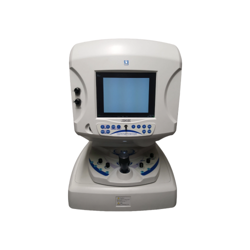
Nidek F-10: High-Precision Retinal and Choroidal Imaging
The Nidek F-10 is a digital laser scanning ophthalmoscope specially developed to provide ophthalmologists with high-definition diagnostic imaging. It ensures clear and detailed observations, even at the capillary level, without the need for image processing after the examination. Thanks to its optimised catadioptric optical system, it captures images of remarkable sharpness, even in the peripheral areas of the retina, with minimal aberrations. Examinations can include infrared images, fluorescein angiography (FA), ICG angiography (IA) or simultaneous angiography, offering maximum flexibility for early detection of pathologies.
Clinical Versatility and Specialised Techniques
The F-10 offers four laser sources with different wavelengths (490 nm, 532 nm, 660 nm and 790 nm), each allowing different layers of the retina to be explored. The autofluorescence mode facilitates the early diagnosis of dry age-related macular degeneration (AMD) without injections and with optimal patient comfort. Retro mode sensitively detects drusen and cystoid macular oedema, while DCO (Differential Contrast Ophthalmoscopy) improves the visualisation of vessels in pathological areas. The device is also suitable for glaucoma monitoring thanks to 532 nm imaging, which allows analysis of the retinal nerve fibre layer (RNFL).
Optimised Workflow and Data Management
The Nidek F-10 is designed to integrate easily into the clinical workflow. Its NAVIS-Lite software offers intuitive management of still images and videos, accurate recording of patient data and standard measurement functions such as cup/disc ratio. Capturing images or video sequences is quick and easy, with direct access to all controls on the front of the device. Panoramic images, fixation guidance and automatic data transfer to a PC help improve productivity and the quality of clinical follow-up.