• Medical Diagnostic Equipment
  • Surgical and Therapeutic Equipment
  • Medical Furniture
  • Laboratory Equipment
  • Cleaning and Sterilization

Fundus Camera VISUCAM 200 - Good Condition

Brand: Zeiss


The ZEISS VISUCAM 200 is a high-performance, fully integrated fundus camera system designed for high-quality retinal imaging. It offers a wide range of imaging modes, including color photography, red-free, blue, anterior segment imaging, stereoscopic imaging, and fundus autofluorescence (FAF). Its intuitive interface and automated features—such as focus and flash settings—ensure ease of use, even for less experienced users. Ideal for screening conditions like AMD or diabetic retinopathy, the VISUCAM 200 delivers sharp, detailed images that enable fast and reliable diagnoses.

Offer Details

VISUCAM® 200 feature legendary ZEISS optics and non-mydriatic color fundus photography enabling you to photograph through pupils as small as 3.3mm. Superior patient comfort, more efficient workflow and improved eye care: The advantages of the VISUCAM 2001 from ZEISS are obvious. The high-quality system provides the basic for detailed diagnoses of typical eye diseases like diabetic retinopathy, glaucoma and AMD in a single workstation.Advanced features such as easy stereo image handling and innovative assessment of macular pigment optical density (MPOD) are combined with intelligent auto functions that enable reproducible and intuitive imaging for every single patient eye.

Description

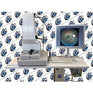
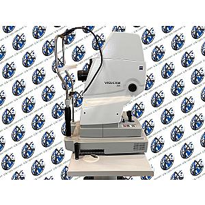
ZEISS VISUCAM 200: High-Precision Retinal Imaging at Your Fingertips

The ZEISS VISUCAM 200 sets a benchmark in fundus imaging by combining all essential ophthalmic evaluation functions in a single compact system. Designed to meet the most demanding clinical requirements, it offers a wide range of imaging modes tailored to various diagnostic needs—including color photography, red-free, blue, anterior segment imaging, fundus autofluorescence (FAF), and stereo capture.

One of the VISUCAM 200’s key strengths lies in its user-centric ergonomics. Automated features such as AutoFocus, AutoFlash, and guided working distance alignment ensure quick, intuitive, and reproducible image acquisition. Even untrained personnel can obtain clinically usable images on their first try. Real-time image display on a 19- or 24-inch flat screen provides excellent visibility, simplifying image review and enhancing patient education.

The integrated FAF technology (Fundus Autofluorescence) enables non-invasive visualization of retinal pigment epithelium (RPE) alterations. This is particularly valuable for the early detection of conditions such as age-related macular degeneration (AMD), hereditary retinal dystrophies, and central serous chorioretinopathy. No contrast agent is required, and the FAF mode offers additional clinical insight into the retina’s metabolic status with ease.

An optional macular pigment density (MPD) module enables automatic, reproducible, and rapid assessments to support AMD risk evaluation. 3D imaging capabilities facilitate longitudinal analysis of glaucomatous changes.

Seamless connectivity with systems like FORUM or VISUPAC ensures efficient image management via network, USB drive, or built-in disc burner. Images can be exported in standard formats (DICOM, JPEG, TIFF, BMP), ensuring broad compatibility and workflow integration.

The ZEISS VISUCAM 200 is a comprehensive, reliable, and modern solution for retinal imaging—designed to enhance diagnostic confidence while simplifying daily clinical operations.

Features

  • High-resolution fundus imaging with automated capture
  • Small pupil mode (≥ 3.3 mm) for difficult-to-image patients
  • FAF imaging for non-invasive analysis of retinal disorders
  • Optional Macular Pigment Density (MPD) measurement module
  • AutoMap function for panoramic retinal montage
  • Real-time display with intuitive navigation
  • Network connectivity and full DICOM compatibility
  • Stereo capture function for 3D imaging
  • Table-mounted system designed for wheelchair accessibility

Technical Details

  • Field of view: 45° and 30°
  • Capture modes: Color, red-free, blue, red, anterior segment, FAF, MPD (optional), stereo
  • Image sensor: 5.0 megapixel CCD
  • Capture time: 1.5 to 2 seconds depending on flash energy
  • Refractive error compensation: Continuously adjustable from +35 D to -35 D
  • Minimum pupil diameter: ≥ 4.0 mm (≥ 3.3 mm in 30° small-angle mode)
  • Working distance: 40 mm (eye to front lens)
  • Flash energy: Xenon flash lamp, 16 levels, max. 30 Ws
  • Monitor: 19" TFT (1280 x 1024) or 24" TFT (1920 x 1200)
  • Computer system: Windows XP Pro, minimum 320 GB hard drive, 1 GB RAM
  • Interfaces: USB ports, network connectors, DVI port
  • Image formats: DICOM-OP, DICOM-VL, DICOM-MWL, BMP, TIFF, JPEG
  • Built-in disc burner: DVD/CD, UDF format
  • Device dimensions: 410 x 480 x 650 mm
  • Weight: 30 kg
  • Nominal voltage: 100–240 V ±10%
  • Power consumption: 400 VA

Compatible Accessories

  • USB printer
  • USB flash drive
  • Sliding keyboard tray
  • FORUM or VISUPAC module for image archiving and analysis
  • Network isolator

Discover more in this category

Logistics

Remma and its partners provide you with serviced and configured equipment tailored to your needs. Receive your product at your facility with standard or express delivery, and an installation option, all through a single point of contact.

Warranty

Our 12/24-month warranty ensures the safety and reliability of your medical equipment throughout its lifespan.

Flexible Financing

Settle your medical equipment in up to 36 installments with our financing service or pay after delivery with Remma Facility.