iCAM: a Reliable Medical Solution for Fundus Documentation
The iCAM from Optovue is a non-mydriatic fundus camera designed to meet the demands of modern ophthalmic practice. With a 45° field of view, it allows the retina, optic disc and anterior segment to be documented with high precision. The device offers two imaging modes: colour capture and red-free imaging, accessible at the touch of a button. This dual capability is particularly useful for clinical evaluation, as colour images allow visualisation of the overall structure and anatomical features, while red-free mode facilitates the assessment of vascular details and structural contrasts. This versatility makes the iCAM suitable for screening examinations, monitoring chronic conditions such as diabetic retinopathy or glaucoma, and standard medical documentation.
High-Precision Clinical Images for Reliable Diagnosis
At the heart of the system is a 12-bit CCD sensor, which offers superior colour fidelity and significantly reduced noise compared to more traditional 8-bit CMOS sensors. This technology provides twenty times greater tonal richness, allowing subtle variations in pigmentation or structure in the ocular tissues to be finely differentiated. With a resolution of 5.2 megapixels, the iCAM guarantees images that can be used for documentation, differential diagnosis and clinical follow-up. An integrated deconvolution algorithm helps improve image sharpness, enhancing the practitioner's ability to detect and analyse retinal abnormalities. The use of an LED flash, which is more durable and energy-efficient than xenon tubes, provides uniform illumination while minimising patient discomfort. These features combine to ensure consistent, reliable image quality, which is essential for clinical ophthalmological assessment.
A Compact, Ergonomic Device Designed for Everyday Use
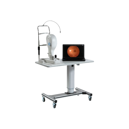
In addition to its imaging performance, the iCAM stands out for its ease of use and portable format. Ergonomics are optimised thanks to an intuitive joystick control that facilitates positioning and focusing. The internal mounting system, complemented by an adjustable external mounting point, ensures stable and comfortable alignment of the patient's gaze. Manual focusing, combined with visual aids, allows rapid adaptation to different patient profiles, even with dioptric variations ranging from -35D to +30D. The software interface is designed to be clear and accessible, reducing operator training time and simplifying the integration of images into electronic medical records (EMR). Compact and lightweight, the device can be installed in various clinical environments, including ophthalmology practices, screening centres and hospitals. Its design makes it a practical tool that combines diagnostic efficiency, ease of use and high-quality medical documentation.