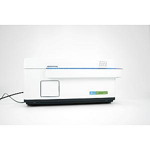
The Operetta: An Solution for Automated Cell Imaging
The Operetta is an automated fluorescence imaging system designed to meet the demands of biomedical research laboratories and cell analysis centers. Developed by PerkinElmer, this cutting-edge device enables HCS (High Content Screening) analyses with exceptional precision, flexibility, and ease of use. Thanks to its advanced integration of imaging technologies, the Operetta is an ideal solution for researchers looking to improve the quality and efficiency of their cellular studies.
The system relies on a 300W xenon lamp, providing a continuous light spectrum from 360 nm to 640 nm. This broad spectral coverage allows for the excitation of a wide range of fluorophores, making the device compatible with various applications in cell imaging and molecular biology. Unlike traditional illumination sources, the xenon lamp ensures uniform intensity, reducing artifacts and improving experimental reproducibility.
One of the Operetta's major advantages is its infrared laser autofocus, which ensures fast and ultra-precise focusing. This technology optimizes image sharpness, even in the presence of complex samples or suspended cells. Coupled with a Peltier-cooled CCD camera (14-bit), this autofocus enables high-resolution image acquisition, essential for morphological and quantitative cell analysis.
The Operetta is designed to adapt to various microplate formats, including 96-well and 384-well plates. Its modular design also allows for the use of custom formats, offering researchers great flexibility. Its optical system, including a range of interchangeable 10X, 20X, 40X, and 60X objectives, ensures optimal observation of cellular structures, from the gross to the subcellular level. For applications requiring higher resolution and reduced background noise, the Operetta also offers an optional confocal mode, allowing for higher-contrast and more detailed images.
The device is fully controlled by Harmony High Content Imaging and Analysis software, which centralizes all operations, from image acquisition to analysis and archiving. Thanks to its intuitive interface, this software facilitates the management of experimental protocols and the interpretation of results, thus reducing the time spent on manual analysis.
Another key advantage of the Operetta is its integration with automation solutions, such as the Plate::Handler II robot. This module enables autonomous microplate management, eliminating the need for human intervention for loading and unloading samples. This automation not only increases laboratory efficiency but also ensures optimal standardization of experimental conditions, thus reducing the risk of errors and improving the reliability of results.
The Operetta stands out for its robustness, ease of use, and versatility, making it an essential tool for cell imaging researchers. Whether for toxicology studies, drug screening, phenotypic analysis, or cell dynamics, this equipment meets the highest standards of precision and efficiency. Combining technologies and an intuitive software interface, the Operetta represents a strategic investment for any laboratory looking to optimize its fluorescence analyses and improve its cell imaging capabilities.


