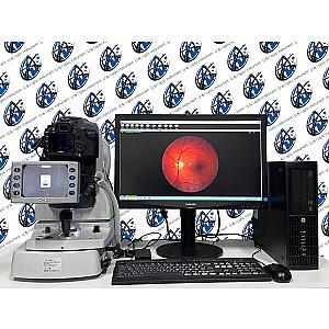
NIDEK AFC-210 Non-Mydriatic Auto Fundus Camera: Advanced Imaging for Effortless Retinal Screening
NIDEK introduces the AFC-210 non-mydriatic auto fundus camera, a cutting-edge solution for effortless retinal screening. This innovative device seamlessly integrates all essential functions required for comprehensive medical examinations, enhancing both the quality and efficiency of the screening process. By incorporating customized built-in functions, the AFC-210 ensures optimal performance and reliability, making it an indispensable tool in clinical practice.
At the heart of the AFC-210 lies its revolutionary imaging optical system, which enables high-resolution digital fundus imaging with unparalleled clarity and detail. This advanced technology delivers precise visualization of the entire fundus, from the bright optic disc to areas affected by retinal pathologies. With significantly reduced noise levels, the system minimizes flash exposure during retinal photography, ensuring quick and efficient image capture while minimizing patient discomfort.
Equipped with an advanced optical system featuring a large sensor, the AFC-210 offers a wide 45° field of view, enabling comprehensive coverage of the retina with exceptional image quality. NIDEK's state-of-the-art auto-tracking technology further enhances the device's functionality, facilitating easy and accurate capture of the anterior corneal center without manual intervention.
The AFC-210 also features an intelligent auto-focus system that seamlessly transitions between anterior and retinal imaging modes, ensuring optimal focusing performance without the need for manual adjustments. Additionally, the inclusion of the unique auto-chinrest feature enhances patient comfort and positioning during examinations.
Complementing its advanced hardware capabilities, the AFC-210 comes equipped with NAVISLite, a sophisticated and user-friendly data filing software. This intuitive software solution streamlines patient data management, allowing healthcare professionals to organize and access patient information with ease, further optimizing workflow efficiency.
In addition to its exceptional imaging capabilities, the AFC-210 offers a range of advanced features to streamline workflow and enhance diagnostic accuracy. Upon image capture, the system automatically sorts images by patient name, simplifying data organization and retrieval. Integrated easy-care pathway protocols ensure seamless display of patient information, facilitating efficient patient management and continuity of care.
The AFC-210 boasts sophisticated imaging functions, including Image Processing, Drawing, Measurement, and Panoramic Imaging, catering to diverse clinical needs. Users can leverage these features to enhance image quality, perform precise measurements, and analyze large field images for comprehensive assessment.
The camera's zoom functionality enables users to magnify images freely, allowing for detailed examination of specific regions of interest. Additionally, a variety of effects such as Sharp, Combination, and Edge Enhancement can be applied to optimize image visualization.
Color control options, including Grey Scale, Contrast RGB, and Red-Free imaging, provide further customization to suit different diagnostic requirements. Users can also rotate or reverse images at any angle, ensuring optimal orientation for analysis and documentation.
Measurement tools, such as C/D Ratio, Disc HV, and Selected Occlusion Area, facilitate quantitative assessment of retinal features, enhancing diagnostic precision. Furthermore, users can insert text or objects directly onto images, enabling annotation for documentation and communication purposes.
The AFC-210 offers flexible print layout display for patient reports, allowing for customized reporting formats tailored to individual preferences or clinical protocols. Additionally, a data backup function ensures data security and integrity, while images can be easily exported for further analysis or archival purposes.
For seamless communication and data sharing, the AFC-210 includes an email function for sending images with accompanying message text. Moreover, the system supports DICOM 3.0 compatibility, enabling seamless integration with DICOM-compatible servers for standardized data exchange and interoperability within healthcare systems. These comprehensive features make the AFC-210 a versatile and indispensable tool for modern ophthalmic practice, facilitating efficient diagnosis, documentation, and management of ocular conditions.