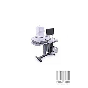
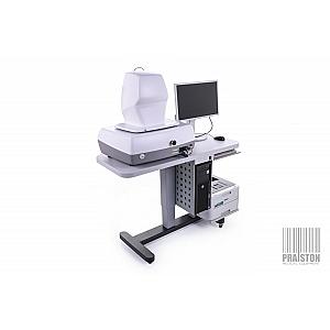
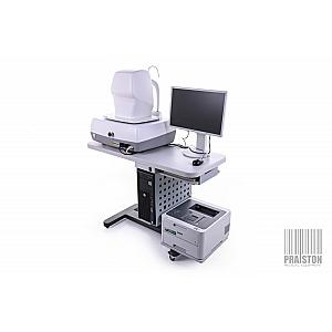
The SOCT Copernicus+ by Optopol is a spectral-domain optical coherence tomography (SOCT) system providing high-resolution images for precise analysis of the retina and glaucoma. With a scanning speed of 27,000 A-scans per second and an axial resolution of 5 µm, it enables rapid and reliable evaluation of ocular structures while ensuring optimal ergonomics for users. Designed to meet the demands of ophthalmologists, this device facilitates the diagnosis and monitoring of retinal and glaucomatous pathologies.
Tomography System SOCT Copernicus+ - Perfect Condition


Offer Details
Accessories :
User manual in Polish (PDF), Power cord, Wired mouse. Wired keyboard, OPTOPOL ACA 160 anterior segment lens,
Seller Warranty :
6-months for domestic market (Poland), 3-months for the international market,
Description
SOCT Copernicus+: For Precise Ocular Analysis
The SOCT Copernicus+ by Optopol is ideal in the field of medical optics, particularly for the analysis of retinal and glaucomatous pathologies. This spectral-domain optical coherence tomography (SOCT) system combines excellent performance in terms of scanning speed, resolution, and ergonomics, providing ophthalmologists and optometrists with a fast, reliable, and precise diagnostic tool.
Designed to meet the demands of ophthalmic practices, the SOCT Copernicus+ utilizes a scanning speed of 27,000 A-scans per second, allowing for extremely rapid image acquisition, which reduces examination time while maintaining optimal image quality. This high scanning speed is complemented by an axial resolution of 5 µm, ensuring highly precise images of the various ocular structures. With this combination, the system enables the visualization of the finest details of the retina and other ocular structures, which are essential for quality diagnostics.
One of the key features of the SOCT Copernicus+ is its ability to perform detailed scans of the retina and optic nerve with clarity. The system allows practitioners to analyze not only the retinal thickness but also the thickness of the retinal nerve fiber layer (RNFL) and the deformation of the retinal pigment epithelium (RPE). This precision is crucial for the early detection of retinal diseases such as macular degeneration and diabetic retinopathy, as well as for monitoring glaucoma. The ability to accurately observe these structures greatly facilitates the detection of subtle changes and can prevent irreversible damage.
The SOCT Copernicus+ also offers several examination modes, including 3D retinal visualization and the Asterisk option for specific analysis of certain structures. In addition to conventional analyses, the device allows vitreoretinal and chorioretinal imaging, providing a comprehensive view of the internal structures of the eye. This is particularly useful when assessing complex pathologies, where a 3D view helps to better understand the relationships between different retinal layers and other ocular structures.
Another major advantage of the SOCT Copernicus+ is its glaucoma module, designed to assist in the detection and management of glaucoma. The module includes the Disk Damage Likelihood Scale (DDLS), a scale for evaluating optic nerve damage. Unlike traditional methods that rely on the cup/disc ratio, the DDLS uses the rim/disc ratio and takes the size of the optic disc into account, thus eliminating the variable effects of disc size. This allows for a more accurate assessment of glaucomatous damage and serves as an essential indicator for tracking the progression of the disease. The SOCT Copernicus+ is therefore a powerful tool for ophthalmologists, as it not only enables the early detection of glaucoma but also allows effective monitoring of disease progression over time.
In terms of ergonomics and ease of use, the SOCT Copernicus+ stands out with its intuitive user interface and One Click functionality, which allows practitioners to quickly start an examination with just a single click. This feature simplifies the examination process and makes the device particularly user-friendly, reducing the risk of errors and enhancing practice efficiency. The system can also be integrated with a centralized server, allowing for easy storage and access to examination data from multiple workstations. This capability is particularly beneficial in clinics and hospitals where data needs to be shared across various practitioners.
Features
- Examination Modes: The SOCT Copernicus+ offers several examination modes, including B-scan, 3D visualization, Asterisk, Animation Scan, and User-defined Scan. These options provide great flexibility in analyzing ocular structures based on the specific needs of the examination.
- Glaucoma Module: The glaucoma module of the SOCT Copernicus+ includes advanced features for glaucoma monitoring and management. This includes the analysis of retinal nerve fiber layer (RNFL) thickness and the evaluation of disease progression. The system uses the Disk Damage Likelihood Scale (DDLS), an innovative scale that analyzes optic nerve damage by considering the disc size and rim morphology.
- Anterior Segment Module: The anterior segment module allows for high-resolution (3 microns) imaging of the cornea and anterior segment. This module is used for measurements such as pachymetry (corneal thickness), LASIK flap thickness, and anterior angle assessment, which is crucial for detecting abnormalities related to angle-closure glaucoma.
- Blood Vessel Recognition: The SOCT Copernicus+ features a highly precise blood vessel recognition system, improving image alignment and ensuring more reliable and reproducible results.
- 3D and Fovea: The advanced 3D module enables visualization of 3D reconstructions of retinal structures, facilitating detailed analysis of local pathologies. It also allows for the visualization of vitreomacular traction, a useful feature for examinations related to macular disorders.
- Layer Recognition System: The new layer recognition system enhances the device’s ability to precisely identify and measure the different layers of the retina and other ocular structures, providing a detailed analysis.
Technical Details
- Scanning speed: 27,000 A-scans per second
- Axial resolution: 5 µm
- Transversal resolution: 12-18 µm
- Axial scanning window width: 2 mm
- B-scan width: 10 mm
- Minimum pupil diameter for measurement: 3 mm
- Light source: 840 nm, 50 nm bandwidth
- Dimensions (H x W x D): 640 x 680 x 520 mm
- Voltage: 230 V, 50 Hz / 115 V, 60 Hz
- Examination modes: B-scan, 3D, Asterisk, Animation Scan, User-defined Scan
- Glaucoma module: DDLS, RNFL thickness analysis, and progression
- Anterior segment module: Pachymetry, LASIK flap, anterior angle measurement
Compatible Accessories
- Review Station: Allows viewing and analyzing captured images.
- CD/DVD: For archiving examination reports.
- Server Module: To centralize and access examination data from multiple workstations.
- Blood Vessel Recognition Software: Enhances the accuracy of retinal analysis.
- Progression and Comparison Module: An essential tool for longitudinal patient monitoring.